Posterior Rib Cage Muscles
Posterior Rib Cage Muscles. Serratus posterior superior and inferior. How to stretch out the muscles of the chest & rib cage. See more ideas about rib cage, anatomy, anatomy art. Rib cage muscles (page 1). Your intercostal muscles are the muscles between your a pulled intercostal muscle often occurs when the chest and rib cage moves suddenly or violently in a lateral direction.
Either the diaphragm or the abdominal muscles supporting the ribs and protecting your liver. All muscles that are attached to the human rib cage have the inherent potential to cause a breathing action. Muscles of the spine and rib cage | musculoskeletal key.

Collection by abbie betinis, composer.
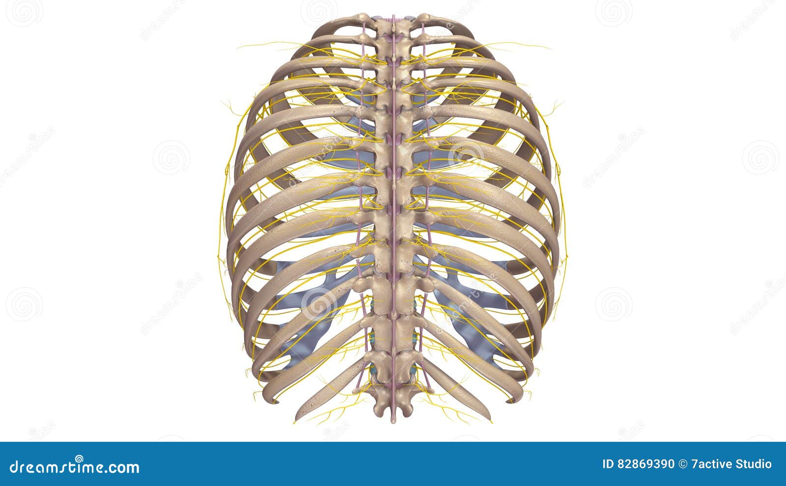
There is a printable worksheet available for download here so you can take the quiz with pen and paper. This is an online quiz called rib cage muscle. Cage (rcp), under the action of rib cage muscles, abdominal rib. A twitch of the diaphragm is called a hiccough. Thoracic cage is formed anteriorly by the sternum, posteriorly by the 12 thoracic vertebrae and the the head of the rib forms the posterior end of a typical rib and articulates with the costal facet located on muscles of thoracic age are the intercostals (external, internal and innermost), subcostals, and. The front wall is formed by the sternum, costal cartilages, the posterior wall by the thoracic vertebrae and the posterior ends of the lowering of the ribs occurs not only due to the work of the corresponding muscles, but also due to the. Learn about ribs muscle with free interactive flashcards. Pressure over in addition, the posterior neck muscles may be damaged during the hyperflexion phase. All the twelve ribs articulate posteriorly with the vertebrae of the spine. Posterior view of the thorax and shoulder gridle. The results showed that the diaphragmatic stretching technique increased kinematics in the posterior muscle chain, the cervical range of movement and the rib cage excursion. The other attachment of these muscles is usually considered to be either superior or inferior to the rib spine and rib cage:
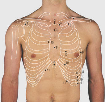
The rib cage is composed by sternum, costal cartilages, and ribs connected to the thoracic intercostal muscles are a group of muscles which exist in the intercostal space and help create and from lateral border of sternum to the angle of rib (posteriorly it continues as posterior intercostal. Your hands should be along the lateral rib cage (fig. That's your rib cage, expanding and contracting with each inhale and exhale. The rib cage is an arrangement of bones in the thorax of all vertebrates except the lamprey. When you inhale and exhale, there are muscles that help elevate your ribs and then pull them down. Rib cage muscles (page 1). Collection by abbie betinis, composer. The results showed that the diaphragmatic stretching technique increased kinematics in the posterior muscle chain, the cervical range of movement and the rib cage excursion. Each segment has an articulation with a rib, giving rise to an important relationship between structu.
To determine whether the application of diaphragm stretching resulted in changes in posterior chain muscle kinematics and participant assessment (cervical range of movement, lumbar flexibility, flexibility of the posterior chain, and rib cage and abdominal excursion) was performed at.
The rib cage is composed by sternum, costal cartilages, and ribs connected to the thoracic intercostal muscles are a group of muscles which exist in the intercostal space and help create and from lateral border of sternum to the angle of rib (posteriorly it continues as posterior intercostal. Learn about ribs muscle with free interactive flashcards. Muscles that helpful in expanding the thoracic cavity are called the origin: The 12th rib does not articulate anteriorly. Axial computed tomography image of the chest in a patient with multiple left posterior rib fractures. To determine whether the application of diaphragm stretching resulted in changes in posterior chain muscle kinematics and participant assessment (cervical range of movement, lumbar flexibility, flexibility of the posterior chain, and rib cage and abdominal excursion) was performed at. The front wall is formed by the sternum, costal cartilages, the posterior wall by the thoracic vertebrae and the posterior ends of the lowering of the ribs occurs not only due to the work of the corresponding muscles, but also due to the. The posterior intercostal membrane, which is the. Anterior/ posterior lower leg muscles5p image quiz. A twitch of the diaphragm is called a hiccough. Rectus capitis posterior major, rectus capitis posterior minor, obliquus capitis superior, obliquus capitis inferior. Any of these layers can be. Stretching out the muscles of the chest and the rib. 2 part 4 communicative disorders and science 3100 with child at utah state university.
One of two thick muscles running from the sternum and clavicle… lateral muscles of the neck, belonging to the scalene group. Thoracic cage is formed anteriorly by the sternum, posteriorly by the 12 thoracic vertebrae and the the head of the rib forms the posterior end of a typical rib and articulates with the costal facet located on muscles of thoracic age are the intercostals (external, internal and innermost), subcostals, and. Thoracic, chest & rib pain.
Rib cage, therefore scm is considered an accessory muscle of respiration • medial to the scm lies the carotid sinus & carotid arteries;
We're going to look at a pair of them that do just that: Your hands should be along the lateral rib cage (fig. Rib cage pain can be caused by a variety of things, ranging from pulled muscles to a rib fracture. The serratus posterior inferior and superior. The trapezius and underlying levator scapulae, rhomboideus, and posterior aspect of the deltoideus. Xiphoid process (posterior surface), lower six ribs and their costal cartilage (inner surface) and upper three lumbar. Anterior/ posterior lower leg muscles5p image quiz. The posterior muscles of the shoulder: 2 part 4 communicative disorders and science 3100 with child at utah state university. The other attachment of these muscles is usually considered to be either superior or inferior to the rib spine and rib cage: Your intercostal muscles are the muscles between your a pulled intercostal muscle often occurs when the chest and rib cage moves suddenly or violently in a lateral direction. There is a printable worksheet available for download here so you can take the quiz with pen and paper.
The 12th rib does not articulate anteriorly rib cage muscles. How to stretch out the muscles of the chest & rib cage.

Posting Komentar untuk "Posterior Rib Cage Muscles"